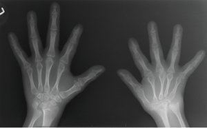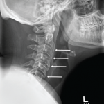
Figure 1: An X-ray of the hands, showing extensive erosion over the ulnar styloid, carpal bones and the carpometacarpal joints of the right side (A), along with abnormal shortening and thinning of diaphysis over the right second through fourth metacarpal bones (B). The X-ray of the left hand appears normal.
Down syndrome (trisomy 21) is one of the most common chromosomal abnormalities. According to the Genomic Resource Centre of the World Health Organization, each year 3,000–5,000 children are born with this chromosome disorder, and about 250,000 families have at least one member with Down syndrome in the U.S.
Down syndrome is caused by numerical aneuploidy, in which the child is born with an extra copy of chromosome 21. This chromosomal aberration affects various systems of the body, mainly neurological, cardiac, endocrinal, gastrointestinal and skeletal.
In this article, we discuss a case of trisomy 21 with a special emphasis on its effect on musculoskeletal health.
Case Presentation
A 9-year-old Caucasian female with Down syndrome was referred to the pediatric rheumatology clinic at Dartmouth in 2011, with complaints of bilateral lower extremity joint pain that had started about two years before. She had no associated history of fever, rashes or vision changes. On examination her joints were hypermobile but without active synovitis or tenderness. Her labs were negative (or normal) for rheumatoid factor (RF), erythrocyte sedimentation rate (ESR), anti-nuclear antibodies (ANA) and C-reactive protein (CRP), and she had a normal thyroid stimulating hormone (TSH) result. Her past medical history was significant for repaired congenital heart disease (bicuspid aortic valve with hypoplastic aortic arch and co-arctation of the aorta), hypothyroidism (controlled with thyroid hormone replacement therapy) and developmental delay. She had no significant family or social history. Because she had no other signs of inflammation, her joint pain was thought to be secondary to her hypermobility, and she was prescribed ibuprofen on as-needed basis and asked to follow up in three months.
Three years later, in June 2014—after missing many follow-up appointments—she came to the clinic again, this time with a six-month history of pain involving the left hip, left foot, cervical spine (C-spine) and interphalangeal joints of both hands, along with right big toe swelling secondary to an injury, but with no evidence of fracture.
On examination, she presented with swelling and tenderness over the interphalangeal joints of the hands and decreased flexion and extension of the C-spine. Again, she had a negative ANA, negative human leukocyte antigen (HLA) B27 and a negative RF. But her ESR was elevated at 23 mm/hr (normal: 0–20 mm/hr), and her CRP was elevated at 7.1 mg/L (normal: <3 mg/L).

